|
R&D Systems
rat anti him trem2 Rat Anti Him Trem2, supplied by R&D Systems, used in various techniques. Bioz Stars score: 93/100, based on 1 PubMed citations. ZERO BIAS - scores, article reviews, protocol conditions and more https://www.bioz.com/result/rat anti him trem2/product/R&D Systems Average 93 stars, based on 1 article reviews
rat anti him trem2 - by Bioz Stars,
2026-03
93/100 stars
|
Buy from Supplier |
|
Bio-Techne corporation
rat anti trem2 Rat Anti Trem2, supplied by Bio-Techne corporation, used in various techniques. Bioz Stars score: 95/100, based on 1 PubMed citations. ZERO BIAS - scores, article reviews, protocol conditions and more https://www.bioz.com/result/rat anti trem2/product/Bio-Techne corporation Average 95 stars, based on 1 article reviews
rat anti trem2 - by Bioz Stars,
2026-03
95/100 stars
|
Buy from Supplier |
|
R&D Systems
anti trem 2 Anti Trem 2, supplied by R&D Systems, used in various techniques. Bioz Stars score: 93/100, based on 1 PubMed citations. ZERO BIAS - scores, article reviews, protocol conditions and more https://www.bioz.com/result/anti trem 2/product/R&D Systems Average 93 stars, based on 1 article reviews
anti trem 2 - by Bioz Stars,
2026-03
93/100 stars
|
Buy from Supplier |
|
R&D Systems
apc anti human trem2 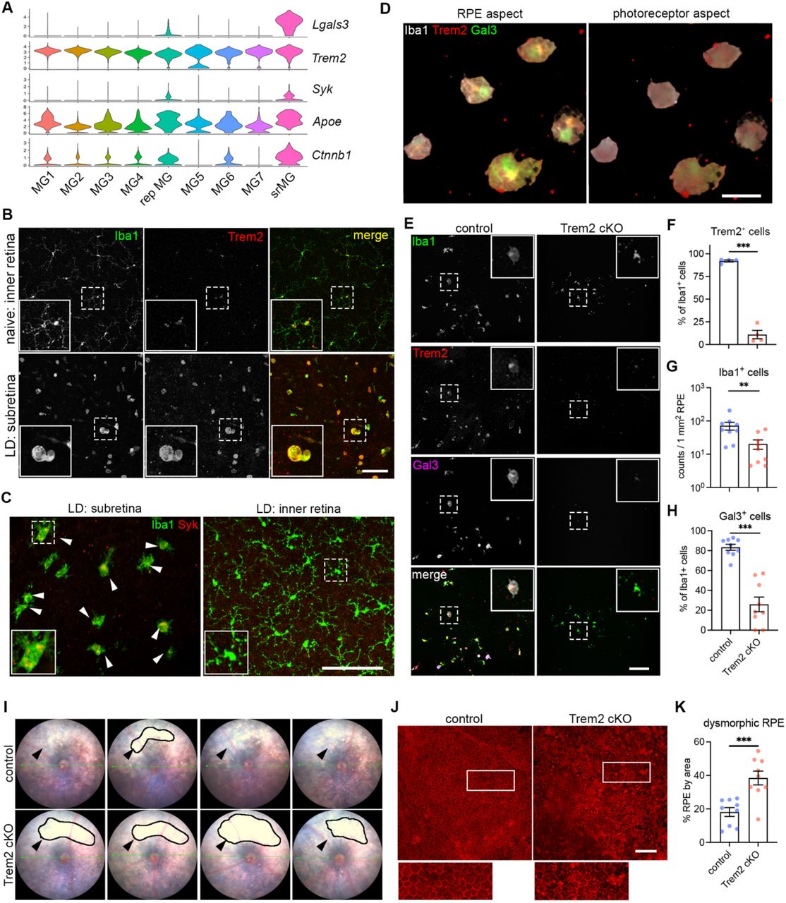 Apc Anti Human Trem2, supplied by R&D Systems, used in various techniques. Bioz Stars score: 93/100, based on 1 PubMed citations. ZERO BIAS - scores, article reviews, protocol conditions and more https://www.bioz.com/result/apc anti human trem2/product/R&D Systems Average 93 stars, based on 1 article reviews
apc anti human trem2 - by Bioz Stars,
2026-03
93/100 stars
|
Buy from Supplier |
|
R&D Systems
anti trem2 Anti Trem2, supplied by R&D Systems, used in various techniques. Bioz Stars score: 93/100, based on 1 PubMed citations. ZERO BIAS - scores, article reviews, protocol conditions and more https://www.bioz.com/result/anti trem2/product/R&D Systems Average 93 stars, based on 1 article reviews
anti trem2 - by Bioz Stars,
2026-03
93/100 stars
|
Buy from Supplier |
|
R&D Systems
trem2 pe  Trem2 Pe, supplied by R&D Systems, used in various techniques. Bioz Stars score: 93/100, based on 1 PubMed citations. ZERO BIAS - scores, article reviews, protocol conditions and more https://www.bioz.com/result/trem2 pe/product/R&D Systems Average 93 stars, based on 1 article reviews
trem2 pe - by Bioz Stars,
2026-03
93/100 stars
|
Buy from Supplier |
|
Proteintech
rabbit anti trem2 Rabbit Anti Trem2, supplied by Proteintech, used in various techniques. Bioz Stars score: 95/100, based on 1 PubMed citations. ZERO BIAS - scores, article reviews, protocol conditions and more https://www.bioz.com/result/rabbit anti trem2/product/Proteintech Average 95 stars, based on 1 article reviews
rabbit anti trem2 - by Bioz Stars,
2026-03
95/100 stars
|
Buy from Supplier |
|
R&D Systems
anti trem2 antibody Anti Trem2 Antibody, supplied by R&D Systems, used in various techniques. Bioz Stars score: 93/100, based on 1 PubMed citations. ZERO BIAS - scores, article reviews, protocol conditions and more https://www.bioz.com/result/anti trem2 antibody/product/R&D Systems Average 93 stars, based on 1 article reviews
anti trem2 antibody - by Bioz Stars,
2026-03
93/100 stars
|
Buy from Supplier |
|
Bioss
trem2 polyclonal antibody Trem2 Polyclonal Antibody, supplied by Bioss, used in various techniques. Bioz Stars score: 94/100, based on 1 PubMed citations. ZERO BIAS - scores, article reviews, protocol conditions and more https://www.bioz.com/result/trem2 polyclonal antibody/product/Bioss Average 94 stars, based on 1 article reviews
trem2 polyclonal antibody - by Bioz Stars,
2026-03
94/100 stars
|
Buy from Supplier |
|
Bio-Techne corporation
human/mouse trem2 apc-conjugated antibody Human/Mouse Trem2 Apc Conjugated Antibody, supplied by Bio-Techne corporation, used in various techniques. Bioz Stars score: 99/100, based on 1 PubMed citations. ZERO BIAS - scores, article reviews, protocol conditions and more https://www.bioz.com/result/human/mouse trem2 apc-conjugated antibody/product/Bio-Techne corporation Average 99 stars, based on 1 article reviews
human/mouse trem2 apc-conjugated antibody - by Bioz Stars,
2026-03
99/100 stars
|
Buy from Supplier |
Image Search Results
Journal: bioRxiv
Article Title: Microglia at Sites of Atrophy Restrict the Progression of Retinal Degeneration via Galectin-3 and Trem2 Interactions
doi: 10.1101/2023.07.19.549403
Figure Lengend Snippet: ( A ) Violin plots showing the upregulation of genes ( Lgals3, Syk and Ctnnb1I) related to Trem2 signaling by subretinal microglia from the integrated dataset of all four mouse models. ( B ) Images of Iba1 (green) and Trem2 (red) staining in naïve microglia from inner retina and subretinal microglia in LD. ( C ) Images of Iba1 (green) and Syk (red) staining in subretinal microglia and microglia from inner retina in LD. ( D ) 3D rendering images of Gal3 (green), Trem2 (red) and Iba1 (white) staining in subretinal microglia in LD. Views from both the apical RPE aspect and neuroretina aspect are shown. ( E ) Images of Iba1 (green), Trem2 (red) and Gal3 (magenta) staining in subretinal microglia between control and Trem2 cKO mice in LD. ( F-H ) Quantifications of Trem2 depletion (F, n=4 per group), Iba1 + cells (G, n=9) and Gal3 + cells (H, n=9) between control and Trem2 cKO mice. ( I ) Fundus images showing increased subretinal white lesions in of Trem2 cKO mice in LD as indicated by arrows. Images from four individual mice per group are shown. ( J ) Images of phalloidin staining in RPE tissues from control and Trem2 cKO mice in LD. ( K ) Quantifications of dysmorphic RPE cells between control and Trem2 cKO mice (n=9 per group). Scale bars: 50μm (D); 100μm (B, C E,and J). Data were collected from 2 independent experiments. **: p<0.01; ***: p<0.001. Unpaired Student’s t-test (F-H).
Article Snippet: Samples were also stained with
Techniques: Staining

Journal: bioRxiv
Article Title: Microglia at Sites of Atrophy Restrict the Progression of Retinal Degeneration via Galectin-3 and Trem2 Interactions
doi: 10.1101/2023.07.19.549403
Figure Lengend Snippet: ( A ) Split views of confocal scans showing the colocalization of Trem2 (red) and Gal3 (green) in the subretinal microglia. Lines indicate the RPE-facing and neuroretina (NR)-facing aspects as indicated. ( B ) Fundus images showing increased subretinal white lesions in anti-Trem2 mAb178 treated mice in LD as indicated by arrows. Images of 4 individual mice per group are shown. ( C ) Images of Iba1 (green) and Gal3 (magenta) staining in subretinal microglia between control and mAb178-treated mice in LD. Scale bar: 100 μm. ( D and E ) Quantifications of Iba1 + cells and Gal3 + cells between control and mAb178 (n=8 per group). ( F ) Images of phalloidin staining in RPE flatmounts from control and mAb178 treated mice in LD. Scale bar: 100μm. ( G ) Quantifications of dysmorphic RPE cells between control (n=8) and mAb178 (n=9) treated mice. ( H ) Images of Iba1 (green) and Trem2 (red) in microglia from the inner retina of naïve control and Trem2 cKO mice. Scale bar: 50μm.
Article Snippet: Samples were also stained with
Techniques: Staining

Journal: bioRxiv
Article Title: Microglia at Sites of Atrophy Restrict the Progression of Retinal Degeneration via Galectin-3 and Trem2 Interactions
doi: 10.1101/2023.07.19.549403
Figure Lengend Snippet: ( A ) ELISA of soluble Trem2 (sTrem2) in vitreous fluid and retinal fluid from naïve WT mice, WT and Trem2 cKO mice subjected to LD. ( B ) Fundus images of mice treated with isotype control or 4D9 anti-Trem2 in LD. Four individual mice per group are shown. ( C ) Representative OCT images of mice treated with isotype or 4D9 in LD. ( D ) Quantifications of outer nuclear layer (ONL) thickness by OCT (n=13 per group). ONL thickness was measured at both nasal and temporal sides. ( E and F ) Scotopic a-waves and b-waves of ERG data among mice treated with isotype or 4D9 in naïve or LD setting (n=5 per group). ( G ) Fundus images of Gal3 cKO mice treated with isotype or 4D9 in LD. Four individual mice per group are shown. ( H ) Representative OCT images of Gal3 cKO mice treated with isotype control or 4D9 anti-Trem2 in LD. ( I ) Quantifications of average ONL thickness by OCT between control and Gal3 cKO mice treated with either isotype or 4D9 (n=13 per group). ( J ) Images of phalloidin staining of control and Gal3 cKO RPE treated with isotype or 4D9 in LD. ( K ) Quantifications of dysmorphic RPE cells (n=15, 13, 11 and 13, respectively). Scale bars: 100μm. Data were collected from 2-4 independent experiments. *: p<0.05; **: p<0.01; ***: p<0.001. Unpaired Student’s t-test (F-H). One-way ANOVA with Tukey’s post hoc test (A); two-way ANOVA with Tukey’s post hoc test (D-F, I and K).
Article Snippet: Samples were also stained with
Techniques: Enzyme-linked Immunosorbent Assay, Staining

Journal: bioRxiv
Article Title: Microglia at Sites of Atrophy Restrict the Progression of Retinal Degeneration via Galectin-3 and Trem2 Interactions
doi: 10.1101/2023.07.19.549403
Figure Lengend Snippet: ( A ) Marker expression of all human clusters. hMG, human microglia; mo-MFs, monocyte-derived macrophages; pv-MFs: perivascular macrophages; mo-DCs, monocyte-derived dendritic cells; VSMC, vascular smooth muscle cells. ( B ) Distribution of clusters by neuroretina and RPE/choroid tissues. Cell number of clusters was normalized to the total counts per tissue. ( C ) Pathway enrichment analysis of subretinal microglia with top 200 shared up-regulated genes. Top significant pathways sorted by false discovery and ranked by fold enrichment are shown. ( D ) UMAP plot showing integrated clustering analysis of three independent human AMD datasets. Data are shown with low resolution to reveal major cell types. ( E ) Dot plot showing the marker expression of major macrophage clusters. Cluster 3 is enriched with RHO expression. ( F ) UMAP plots showing the presence of hMG2 cluster in all three scRNA-seq datasets as indicated by arrows. ( G ) UMAP plots showing the enrichment of cluster 3 in donor 0106_nAMD. ( H ) UMAP plots showing clustering analysis with high resolution by each dataset and comparable heterogeneity of microglia (cluster 0, 7 and 12). As dataset GSE183320 does not contain neurosensory retina tissues, few cells of major homeostatic microglia (cluster 0) are observed in this dataset. ( I ) Violin plots showing the expression of LGALS3 , TREM2 and CD68 by microglial clusters between non-AMD and AMD donors. Both cluster 7 and 12 show LGALS3 upregulation as hMG2 cluster identified in this study. ( J and K ) Quantifications of LGALS3 + microglial clusters (7 and 12) in the macular and whole RPE/choroid tissues between non-AMD and AMD donors. Data were from three independent datasets and compared using Mann-Whitney test. P-values are shown. ns: not significant.
Article Snippet: Samples were also stained with
Techniques: Marker, Expressing, Derivative Assay, MANN-WHITNEY

Journal: bioRxiv
Article Title: Microglia at Sites of Atrophy Restrict the Progression of Retinal Degeneration via Galectin-3 and Trem2 Interactions
doi: 10.1101/2023.07.19.549403
Figure Lengend Snippet: ( A ) Multispectral imaging of GAL3 and CD68 co-staining in the subretinal space (top) and inner retina (bottom) from human donors. Unmixed purple spectrum (GAL3) and yellow spectrum (CD68) are shown. The areas of colocalized spectra are highlighted in green. Scale bar: 50μm. ONL and INL, outer and inner nuclear layers. ( B ) Representative image of Gal3 and CD68 co-staining in the macular GA region of a retinal section from an 88-year-old female donor eye with advanced AMD (Sarks V). Black insert box shows the magnification of GA with double positive cells. Scale bar: 200μm. ONL and INL, outer and inner nuclear layers; GCL, ganglion cell layer. ( C ) Correlation between the frequencies of macular Gal3 + CD68 + double positive cells (y axis) and Sarks AMD grading (x axis) by Spearman’s correlation (n = 18 donors, Table S2). Coefficient and p-value are shown. ( D ) Histograms showing increased TREM2 + myeloid cells (CD45 + CD11B + ) in RPE/choroid tissues of AMD donors. Concatenated histograms were shown (n=3 per groups). Control human blood samples were used to set up flow gating. ( E-G ) Quantifications of TREM2 + (E), CD45 + (F), and CD11B + (G) cell frequencies in RPE/choroid tissues between non-AMD and AMD donors. Unpaired Student’s t test is used. P-values are shown. ( H ) Correlation between the frequencies of TREM2 + myeloid cells (y axis) and Sarks AMD grading (x axis) in RPE/choroid tissues by Spearman’s correlation. Coefficient and p-value are shown.
Article Snippet: Samples were also stained with
Techniques: Imaging, Staining

Journal: bioRxiv
Article Title: Microglia at Sites of Atrophy Restrict the Progression of Retinal Degeneration via Galectin-3 and Trem2 Interactions
doi: 10.1101/2023.07.19.549403
Figure Lengend Snippet: ( A ) Images of GAL3 (purple) and CD68 (yellow) co-staining in the macula region of retinal sections from human donors categorized by Sark grades (I-VI). The macular neurosensory retinas of some subject eyes exhibited fixation-related artifactual detachment. In these subjects, separate images of RPE/choroid tissues are shown. Scale bar: 100μm. ONL and INL, outer and inner nuclear layers. GCL, ganglion cell layer. ( B ) Spectral imaging of GAL3 and CD68 co-staining in the geographic atrophy from donor #23 with advanced AMD (Sarks V). Unmixed purple spectrum (GAL3) and yellow spectrum (CD68) are shown. The areas of colocalized spectra are highlighted in green. Scale bar: 50μm. ( C and D ) Images showing the presence of subretinal GAL3 (purple) and CD68 (yellow) double positive cells in the areas with photoreceptor loss and preserved RPE in the transitional area of the macula from an AMD donor (C) and in the age-related peripheral degeneration of a non-AMD donor (D). Scale bars: 100μm. ( E ) Gating strategy of flow cytometry analysis. CD45 + CD11B + cells and CD45 + CD11B - cells from control blood were used to determine the gating of TREM2 + cells. Concatenated plots are shown for non-AMD and AMD. ( F ) Flow contour plots of individual donors showing increased percentage of TREM2 + myeloid cells in AMD.
Article Snippet: Samples were also stained with
Techniques: Staining, Imaging, Flow Cytometry
Journal: Journal of Neuroinflammation
Article Title: High-salt diet downregulates TREM2 expression and blunts efferocytosis of macrophages after acute ischemic stroke
doi: 10.1186/s12974-021-02144-9
Figure Lengend Snippet: Excess salt downregulates efferocytic molecule TREM2 in macrophages and impairs their inflammation resolution functions. a Macrophages cultured in a high-salt environment were subjected to a PCR array to analyze the expression of efferocytosis-associated receptors in vitro. Data are displayed as fold change relative to macrophages from the PBS-treated group. Experiments were repeated three times. ** P < 0.01 versus PBS group in t test. b , c Protein expression of TREM2 in macrophages after HS treatment was evaluated with western blot ( b ) and flow cytometry ( c ) in vitro. Experiments were repeated three times. * P < 0.05 versus PBS-treated group in t test. d – g ND and HSD mice were subjected to 60min of tMCAO. The brains were collected at 3 days after tMCAO. d mRNA expression of Trem1 and Trem2 in the ipsilateral hemisphere of stroke mice was analyzed with RT-PCR in vivo. CT value was normalized to that of ND mice. N = 3 mice per group. * P < 0.05 versus ND group in t test. e Protein expression of TREM2 in the ipsilateral hemisphere of stroke mice with western blot. N = 6 mice per group. * P < 0.05, versus ND group in t test. f In vivo, TREM2 protein expression in infiltrated macrophages (CD45 hi CD11b + Ly6G - ) of stroke mice was analyzed with flow cytometry. N = 4 mice per group. * P < 0.05 versus ND group in t test. g In vivo CD16 and CD206 expression in TREM2 lo and TREM2 hi macrophages (CD45 hi CD11b + Ly6G - ) in the ipsilateral hemisphere from ND and HSD mice was measured via flow cytometry. The mean fluorescence intensity (MFI) of CD16 or CD206 was quantified. N = 4 mice per group. * P < 0.05 and ** P < 0.01 versus TREM2 lo macrophages in t test
Article Snippet: The following antibodies were used: CD45-PE-Texas Red (1:400, BioLegend), CD11b-PE (1:400, BioLegend), CD3-PerCp/Cy5.5 (1:400, BioLegend), CD19-FITC (1:400, BioLegend), Ly6G-APC/Cy7 (1:400, BioLegend),
Techniques: Cell Culture, Expressing, In Vitro, Western Blot, Flow Cytometry, Reverse Transcription Polymerase Chain Reaction, In Vivo, Fluorescence

Journal: Journal of Neuroinflammation
Article Title: High-salt diet downregulates TREM2 expression and blunts efferocytosis of macrophages after acute ischemic stroke
doi: 10.1186/s12974-021-02144-9
Figure Lengend Snippet: TREM2 expression is decreased in AIS patients with high-salt intake and is correlated with the proinflammatory property of circulating monocytes and detrimental stroke outcomes. AIS patient dietary salt intake was measured with a 24-h urine sodium (24h UNa) with a normal limit of 170 mmol. a Representative images of the MRI-DWI of AIS patients with normal or high urine sodium concentration. Comparison of infarct size of AIS patients with normal ( N = 13) or high ( N = 25) urine sodium concentration. * P < 0.05 by Student’s t test. b Comparison of 1-day NIHSS, 7-day NIHSS of AIS patients with normal ( N = 13) or high ( N = 25) urine sodium concentration. * P < 0.05 by Student’s t test . c Heat map showing TREM2, CD206, and CD80 protein expression in peripheral monocytes of AIS patients with normal ( N = 13) or high ( N = 25) urine sodium concentration using flow cytometry. The function of “scale” was applied for value normalization. d Comparison of CD206 and CD80 protein expression in peripheral monocytes of AIS patients with normal ( N = 13) or high urine sodium concentration ( N = 25) using flow cytometry. * P < 0.05 by Student’s t test. e Comparison of TREM2 protein expression in peripheral monocytes of AIS patients with normal ( N = 13) or high urine sodium concentration ( N = 25) using flow cytometry. * P < 0.05 by Student’s t test . f Comparison of TREM2 mRNA expression in PBMCs of AIS patients with normal ( N = 6) or high urine sodium concentration ( N = 12). * P < 0.05 by Student’s t test. g Correlation of clinic parameters, the protein expression of TREM2 and 24h UNa was assessed with Spearman correlation analysis. * P < 0.05, ** P < 0.01. DWI diffusion-weighted imaging, DM diabetes mellitus, HL hyperlipidemia, CHD coronary heart disease, AF atrial fibrillation, WBC white blood cell, NLR neutrophil-to-lymphocyte ratio, BNa blood sodium, CRP C-reactive protein, HCY homocysteine, and Hba1c glycated hemoglobin
Article Snippet: The following antibodies were used: CD45-PE-Texas Red (1:400, BioLegend), CD11b-PE (1:400, BioLegend), CD3-PerCp/Cy5.5 (1:400, BioLegend), CD19-FITC (1:400, BioLegend), Ly6G-APC/Cy7 (1:400, BioLegend),
Techniques: Expressing, Concentration Assay, Comparison, Flow Cytometry, Diffusion-based Assay, Imaging

Journal: Journal of Neuroinflammation
Article Title: High-salt diet downregulates TREM2 expression and blunts efferocytosis of macrophages after acute ischemic stroke
doi: 10.1186/s12974-021-02144-9
Figure Lengend Snippet: Enhancement of TREM2 signaling restores the efferocytic capacity and cellular inflammation resolution of macrophages in a high salinity environment. Bone marrow-derived primary cultured macrophages were infected with lentiviral vectors carrying TREM2-GFP cDNA (TREM2-Lenti) or control lentivirus carrying GFP only (NC-Lenti). Macrophages were subjected to analysis at 2 days after infection. a , b Transfected macrophages were treated in vitro with HS or PBS overnight and then subjected to efferocytic analysis with PI + dead/dying neurons. The efferocytic efficiency of macrophages was evaluated with immunostaining ( a ) and flow cytometry ( b ). Engulfed dead/dying neurons (PI + Phalloidin + ) per macrophage or the proportion of phagocytic macrophages (PI + F4/80 + ) was calculated at the corresponding time points. Data were collected from three independent experiments. * P < 0.05 and *** P < 0.001 versus NC-Lenti PBS group in one-way ANOVA. c In vitro protein expression of Arg1 and CD206 in transfected macrophages with HS or PBS treatment overnight was analyzed with flow cytometry. * P < 0.05 versus NC-Lenti PBS group in one-way ANOVA. d , e C57BL/6 mice with HSD or ND were first injected with clodronate liposomes to deplete monocytes/macrophages. Mice were then subjected to 60 min of tMCAO. Immediately after reperfusion, a single dose of 2x10 6 TREM2-overexpressing macrophages or NC macrophages were intravenously transferred in vivo. d The in vivo infarct volume of male mice was quantified on NeuN (red)-stained coronal sections at 3 days. Dashed lines outline the infarct area. N = 6 mice per group, * P < 0.05, ** P < 0.01, and **** P < 0.0001, one-way ANOVA . e Confocal microscopy analysis of NeuN (blue) and Iba1 (red) double-staining in HSD mice. Quantification of the total number of Iba1 + NeuN + cells (microglia/macrophages that have engulfed neurons) and phagocytic index (the proportion of neurons engulfed by microglia/macrophages) in ischemic areas of HSD mice. N = 4 mice per group. * P < 0.05 and ** P < 0.01 versus NC macrophage group in t test. f In vivo CD206 (red) expression in Iba1 + GFP + cells was analyzed via immunostaining. N = 4 mice per group. * P < 0.05 versus NC macrophage group in t test
Article Snippet: The following antibodies were used: CD45-PE-Texas Red (1:400, BioLegend), CD11b-PE (1:400, BioLegend), CD3-PerCp/Cy5.5 (1:400, BioLegend), CD19-FITC (1:400, BioLegend), Ly6G-APC/Cy7 (1:400, BioLegend),
Techniques: Derivative Assay, Cell Culture, Infection, Transfection, In Vitro, Immunostaining, Flow Cytometry, Expressing, Injection, Liposomes, In Vivo, Staining, Confocal Microscopy, Double Staining